About the Department of Histology and Embryology
The Department of Histology and Embryology provides a complete range of teaching in the fields of cytology, general histology, functional organ and tissue histology, and embryology for the Master’s degree programme General Medicine and three Bachelor’s degrees (Laboratory Assistant, Midwife, Public Health). The Department’s staff participates in both theoretical instruction and practical training.
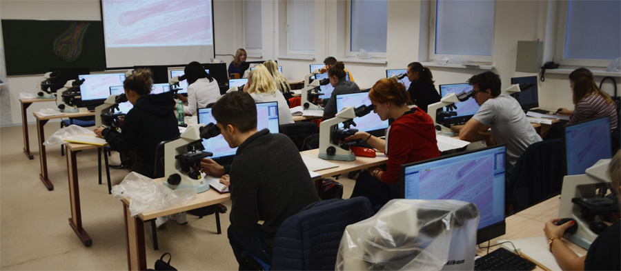
Theoretical instruction (in the form of lectures) covers the histology of tissues, organs and systems, including numerous specific clinical correlations which clearly demonstrate their morphology and functions. The lectures also emphasize the integrity of the human organism and the mutual interrelations among its component systems. The practical tuition takes place in the microscopy hall, with 20 microscopes and audiovisual equipment. Students already know from their anatomy courses what the human body looks like at a microscopic scale; working with microscopes, they view histological specimens and learn to recognize different types of human tissues and cells. The specimens are also captured on the Olympus dotSlide microscope and stored in a database of virtual histological specimens. This represents a major contribution to teaching – students can view different structures on computers before progressing to microscope work. Students can also study histological specimens at home.
Students can expand their knowledge by attending the optional course “Immunohistochemical detection of normal and pathological human tissues”, which gives an introduction to special immunohistochemical methods applied to specific cells and tissues – and the pathological conditions with which they are associated.
The Department is currently in the process of building a new laboratory which will be used to prepare histological specimens for students as well as to conduct standard and immunohistochemical dyeing for research purposes.
Updated: 24. 02. 2022


